The heart is the body's engine room, responsible for pumping life-sustaining blood via a 60,000-mile-long (97,000-kilometer-long) network of vessels. The organ works ceaselessly, beating 100,000 times a day, 40 million times a year—in total clocking up three billion heartbeats over an average lifetime. It keeps the body freshly supplied with oxygen and nutrients, while clearing away harmful waste matter.
The fetal heart evolves through several different stages inside the womb, first resembling a fish's heart, then a frog's, which has two chambers, then a snake's, with three, before finally adopting the four-chambered structure of the human heart.
Function
About the size of its owner's clenched fist, the organ sits in the middle of the chest, behind the breastbone and between the lungs, in a moistened chamber that is protected all round by the rib cage. It's made up of a special kind of muscle (cardiac muscle) that works involuntarily, so we don't have to think about it. The heart speeds up or slow downs automatically in response to nerve signals from the brain that tell it how much the body is being exerted. Normally the heart contracts and relaxes between 70 and 80 times per minute, each heartbeat filling the four chambers inside with a fresh round of blood.
These cavities form two separate pumps on each side of the heart, which are divided by a wall of muscle called the septum. The upper chamber on each side is called the atrium. This is connected via a sealing valve to the larger, more powerful lower chamber, or ventricle. The left ventricle pumps most forcefully, which is why a person's heartbeat is felt more on the left side of the chest.
When the heart contracts, the chambers become smaller, forcing blood first out of the atria into the ventricles, then from each ventricle into a large blood vessel connected to the top of the heart. These vessels are the two main arteries. One of them, the pulmonary artery, takes blood to the lungs to receive oxygen. The other, the aorta, transports freshly oxygenated blood to the rest of the body. The vessels that bring blood to the heart are the veins. The two main veins that connect to the heart are called the vena cava.
Blood Delivery
Since the heart lies at the center of the blood delivery system, it is also central to life. Blood both supplies oxygen from the lungs to the other organs and tissues and removes carbon dioxide to the lungs, where the gas is breathed out. Blood also distributes nourishment from the digestive system and hormones from glands. Likewise our immune system cells travel in the bloodstream, seeking out infection, and blood takes the body's waste products to the kidneys and liver to be sorted out and trashed.
Given the heart's many essential functions, it seems wise to take care of it. Yet heart disease has risen steadily over the last century, especially in industrialized countries, largely due to changes in diet and lifestyle. It has become the leading cause of death for both men and women in the United States, claiming almost 700,000 lives a year, or 29 percent of the annual total. Worldwide, 7.2 million people die from heart disease every year.
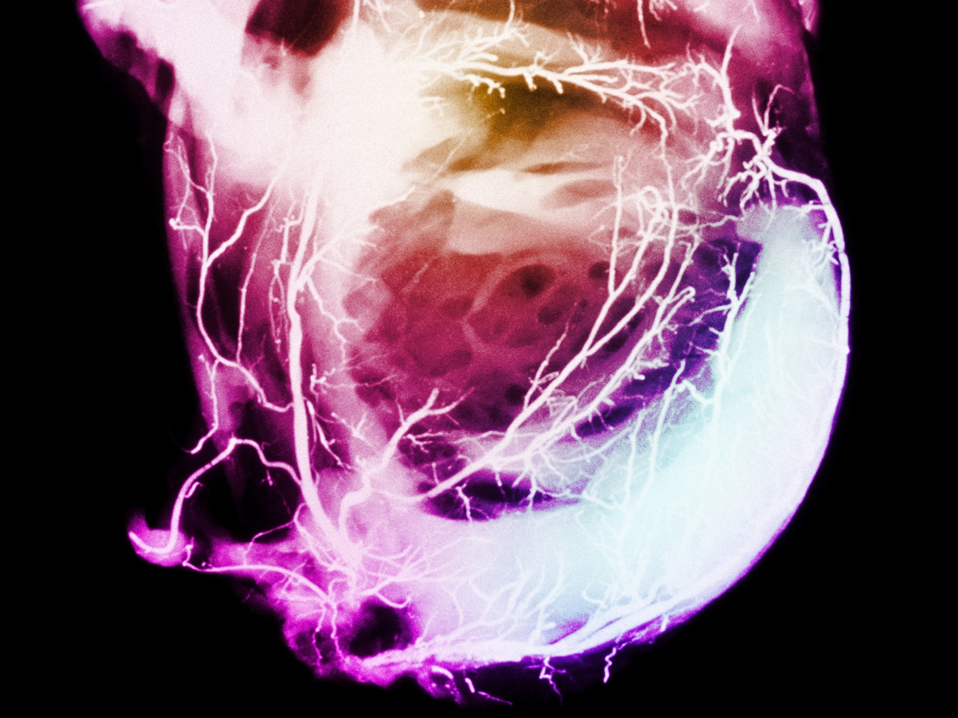
See Photos of Heart Surgery, Patients, and Diseases


You May Also Like


Go Further
Animals
- This ‘saber-toothed’ salmon wasn’t quite what we thoughtThis ‘saber-toothed’ salmon wasn’t quite what we thought
- Why this rhino-zebra friendship makes perfect senseWhy this rhino-zebra friendship makes perfect sense
- When did bioluminescence evolve? It’s older than we thought.When did bioluminescence evolve? It’s older than we thought.
- Soy, skim … spider. Are any of these technically milk?Soy, skim … spider. Are any of these technically milk?
- This pristine piece of the Amazon shows nature’s resilienceThis pristine piece of the Amazon shows nature’s resilience
Environment
- This pristine piece of the Amazon shows nature’s resilienceThis pristine piece of the Amazon shows nature’s resilience
- Listen to 30 years of climate change transformed into haunting musicListen to 30 years of climate change transformed into haunting music
- This ancient society tried to stop El Niño—with child sacrificeThis ancient society tried to stop El Niño—with child sacrifice
- U.S. plans to clean its drinking water. What does that mean?U.S. plans to clean its drinking water. What does that mean?
History & Culture
- Séances at the White House? Why these first ladies turned to the occultSéances at the White House? Why these first ladies turned to the occult
- Gambling is everywhere now. When is that a problem?Gambling is everywhere now. When is that a problem?
- Beauty is pain—at least it was in 17th-century SpainBeauty is pain—at least it was in 17th-century Spain
- The real spies who inspired ‘The Ministry of Ungentlemanly Warfare’The real spies who inspired ‘The Ministry of Ungentlemanly Warfare’
- Heard of Zoroastrianism? The religion still has fervent followersHeard of Zoroastrianism? The religion still has fervent followers
Science
- Here's how astronomers found one of the rarest phenomenons in spaceHere's how astronomers found one of the rarest phenomenons in space
- Not an extrovert or introvert? There’s a word for that.Not an extrovert or introvert? There’s a word for that.
- NASA has a plan to clean up space junk—but is going green enough?NASA has a plan to clean up space junk—but is going green enough?
- Soy, skim … spider. Are any of these technically milk?Soy, skim … spider. Are any of these technically milk?
- Can aspirin help protect against colorectal cancers?Can aspirin help protect against colorectal cancers?
Travel
- What it's like to hike the Camino del Mayab in MexicoWhat it's like to hike the Camino del Mayab in Mexico
- Is this small English town Yorkshire's culinary capital?Is this small English town Yorkshire's culinary capital?
- Follow in the footsteps of Robin Hood in Sherwood ForestFollow in the footsteps of Robin Hood in Sherwood Forest
- This chef is taking Indian cuisine in a bold new directionThis chef is taking Indian cuisine in a bold new direction
